ECM Kits/Liver Fibrotic dECM
Liver Fibrotic dECM
ECM Kits/Liver Fibrotic dECM
Liver Fibrotic dECM
Product Number: MTHDLV201
Size: 10 mL
TissueSpec® Human Liver Fibrotic dECM Coating Kit brings the fibrotic liver matrix environment derived from human livers to your 2D culture. Each lot is specific to 1 donor and is sufficient to prepare 10 mL of coating at a concentration of 0.1 mg/mL. Research use only.
Kit contents
1 x 1 mL TissueSpec® Human Liver Fibrotic dECM (1 mg/mL)
1 x 1 mL 10x Buffer
Material properties
Origin: Liver (human)
Composition: Characterized by mass spectrometry
Stiffness: ##
Usage
Use: 2D cell culture
Preparation temperature: 25ºC
Preparation time: 15 min
Hydrogel volume: 10 mL
Number of wells: 1 x 6 well plate at 1.5 mL per well (calculator)
Storage: 2–8ºC
Shelf life: 12 months
Validated cell types: primary human hepatocytes, hepatocarcinoma cells (HepG2), primary human hepatic stellate cells, breast cancer cells (BT-549, T47-D), fibroblasts
Validated multi-well formats: 6, 12, 24, 48, 96, 384
Validated imaging modalities: Brightfield, phase-contrast, immunofluorescence
Applications: Drug testing, disease modeling, high throughput screening
Quality
Biocompatibility: >95% viability human primary fibroblasts
Sterility: Negative for bacteria, fungus, mycoplasma
Concentration: 0.1 mg/mL (recommended working concentration)
pH: Neutral
Gelation time: N/A
Ordering
Lead time: 5 business days
Shipping temperature: 4ºC (wet ice, natural insulating material)
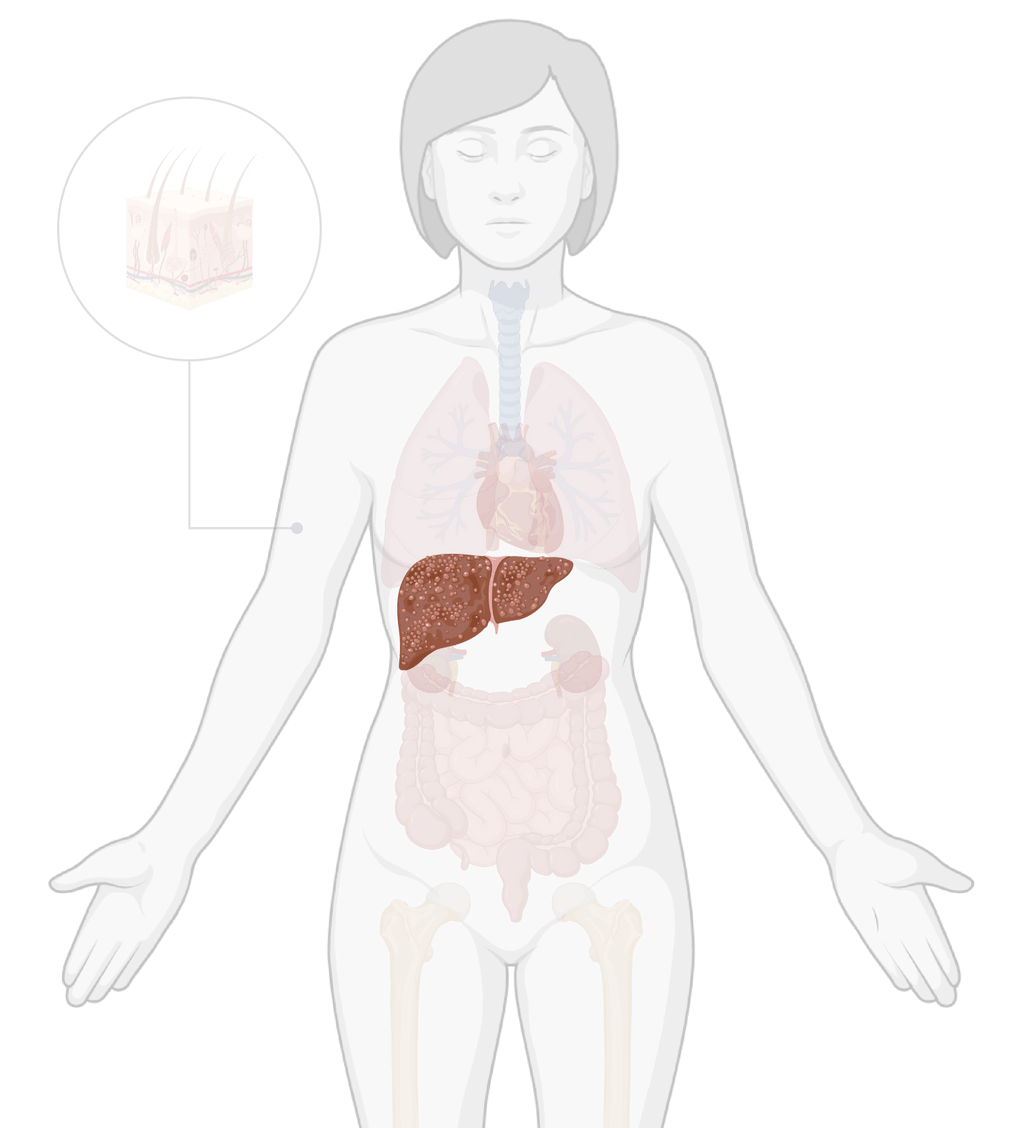
TissueSpec® Human Liver Fibrotic dECM Coating Kit (10 mL)
TissueSpec® Human Liver Fibrotic dECM Coating Kit
TissueSpec® Human Liver Fibrotic dECM Kits bring the fibrotic liver matrix environment derived from human livers to your cell culture.
TissueSpec® Human Liver Fibrotic dECM Coating Kit: Fluorescent labeling of fibrotic liver dECM protein fibers
TissueSpec® Human Liver Fibrotic dECM and TissueSpec® Liver dECM Coating were labeled with Alexa Fluor™ 647 NHS Ester to visualize protein distribution in a 12-well plate and imaged at 20x magnification using the Cy5 channel on a Cytation™ 5 imaging system (Agilent BioTek, USA). Scale bar = 150 µm.
Engineered Human Liver Fibrotic Model: 3D Human Heterospheroids in TissueSpec® Human Liver Fibrotic dECM with overlaid Kupffer cells
Metabolic Activity of engineered human liver fibrotic lesions
Hepatocytes-Hepatic Stellate Cells (HepaRG-HSC) heterospheroids were embedded within healthy and custom TissueSpec® Human Liver Fibrotic dECM hydrogel, and patient-derived Kupffer cells were seeded on top of the gels. Collagen I was used as the control, non-tissue specific substrate. Cultures were treated with 5 ng/mL TGF-β1 over 3 days. Metabolic activity was measured via PrestoBlue™ assay.
TGF-β1 treatments increases CTGF secretion in engineered human liver fibrotic lesions
Hepatocytes-Hepatic Stellate Cells (HepaRG-HSC) heterospheroids were embedded within healthy and custom TissueSpec® Human Liver Fibrotic dECM hydrogel, and Kupffer cells were seeded on top of the gels. Collagen I was used as the control, non-tissue specific substrate. Cultures were treated with 5 ng/mL TGF-β1 over 3 days.
Supernatants were collected and co-cultures were supplemented each day with fresh cell media with or without TGF-β1. CTGF concentrations were quantified by ELISA. Presented values represent the cumulative CTGF secretion per spheroid over the 3-day InMatrico® culture period.
TGF-β1 treatments increases Pro-Collagen I secretion in engineered human liver fibrotic lesions
Hepatocytes-Hepatic Stellate Cells (HepaRG-HSC) heterospheroids were embedded within healthy and custom TissueSpec® Human Liver Fibrotic dECM, and Kupffer cells were seeded on top of the gels. Collagen I was used as the control, non-tissue specific substrate. Cultures were treated with 5 ng/mL TGF-β1 over 3 days.
Supernatants were collected and co-cultures were supplemented each day with fresh cell media with or without TGF-β1. Pro-collagen I concentrations were quantified by ELISA. Presented values represent the cumulative Pro-Collagen I secretion per spheroid over the 3-day InMatrico® culture period.
Morphology of 3D human liver heterospheroids
Representative images of the morphology of heterospheroids (Hepatocytes-Hepatic Stellate Cells (HepaRG-HSC)) embedded within custom TissueSpec® Human Liver Fibrotic dECM hydrogel, healthy liver dECM hydrogel and Collagen I. Kupffer cells (KC) were seeded on top of the gels. Co-cultures were incubated for 3 days with or without 5 ng/mL TGF-β1. Scale bar = 200 µm.